Cardiovascular MRI
diagnosis
table of contents
What is Cardiac MRI?
What does MRI scanner look like
How does the MRI scanner work?
What are some common uses of the procedure? (Indication)
Types of MRI procedures (For various conditions)
How should I prepare?
How is the procedure performed?
How long does the procedure take?
What will I experience during and after the procedure?
What are the risks?
What are the limitations of a Cardiac MRI?
What is Cardiac MRI?
Magnetic resonance imaging (MRI) is a noninvasive medical test that uses a powerful magnetic field, radio frequency pulses and a computer to produce detailed pictures of organs, soft tissues, bone and virtually all other internal body structures. MRI does not use ionizing radiation (x-rays).
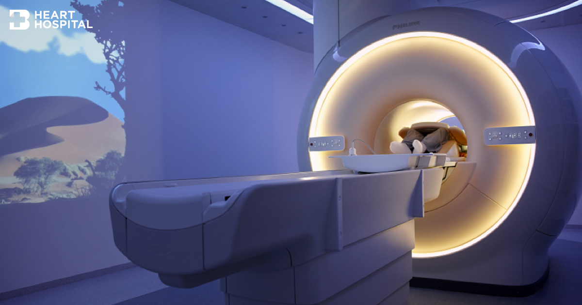
What does MRI scanner look like
Outside
The MRI scanner is on the left. The CT scanner is on the right. They look like giant donut from the front. The MRI scanner is usually deeper, although there are some variations such as short-bore systems, larger diameter bore and open on the sides (open MRI) which is also available at Bangkok heart Hospital.
Inside
The MRI scanner has powerful magnet and radio frequency coil while the CT scanner has X-rays tube at 2 o’clock and CT detectors are on opposite side at 8 o’clock. Only the CT scanner that spins around the patient at very fast spee
How does the MRI scanner work?
Unlike conventional x-ray examinations and computed tomography (CT) scans, MRI does not depend on ionizing radiation. Instead, while in the magnet field, radio waves redirect alignment of hydrogen atoms that naturally exist within the body without causing any chemical changes in the tissues. As the hydrogen atoms return to their usual alignment, they emit energy that varies according to the type of body tissue in which they lie. The MR scanner picks up this energy and creates a picture of the tissues scanned.
The magnetic field is produced by passing an electric current through wire coils in most MRI units. Other coils may be placed around the part of the body being imaged, send and receive radio waves, producing signals that are detected by the coils.
A computer then processes the signals and generates a series of images, each of which shows a thin slice of the body. The images can then be studied from different angles by the interpreting radiologist. Frequently, the differentiation of abnormal (diseased) tissue from normal tissues is better with MRI than with other imaging modalities such as x-ray, CT and ultrasound.
What are some common uses of the procedure? (Indication)
Cardiac MRI imaging is performed to help your physician detect or monitor cardiac disease by:
- Evaluating the anatomy and function of the heart chambers, valves, size and blood flow through major vessels, and surrounding structures such as the pericardium (the sac that surrounds the heart).
- Diagnosing a variety of cardiovascular (heart and/or blood vessel) disorders.
- Evaluating the effects of coronary artery disease such as limited blood flow to the heart muscle and scarring within the heart muscle after a heart attack.
- Planning a patient’s treatment for cardiovascular disorders.
- Monitoring the progression of certain disorders over time.
- Evaluating the anatomy of the heart and blood vessels in children and adults with congenital heart disease. Evaluating the effects of surgical changes, especially in patients with congenital heart disease.
Types of MRI procedures (For various conditions)
- With contrast (gadolinium) injection. Gadolinium contains no iodine.
- Cardiac perfusion study
- Viability study
- MRA angiography
- Without contrast injection
- MR angiography for coronary artery , great vessels and peripheral arteries
- Cardiac function assessment
- Cardiac valvular assessment
- Pericardium and myocardium assessment
- Investigation of specific cardiac pathology ; arrhythmogenic right ventricular dysplasia
- With contrast and drug injection
- MRI stress test with adenosine or dobutamine induction
How should I prepare?
- Wearing: Loose-fitting clothing without metal fasteners, or hospital gown.
- Food/fluid: Depending the area or type of examination. Check with your doctor or nurse. Unless you are told otherwise, you may take medications as usual.
- Do not wear these objects during MRI exam: Metal pins, hairpins, metal zippers, similar metallic items, jewelry and eyeglasses, because they can interfere with the magnetic field of the MRI unit.
- Watches, credit cards and hearing aids and electronic objects all of which can be damaged.
- Removable dental work, body piercings.
- In most cases, an MRI exam is safe for patients with metal implants, except for a few types such as cochlear (ear) implant, some types of clips used on brain aneurysms, some types of metal coils placed within blood vessels and nearly all cardiac defibrillators and pacemakers.
- Some implanted devices require a short period of time after placement (usually six weeks) before being safe for MRI examinations. Examples include but are not limited to: Artificial heart valves, implanted drug infusion ports, artificial limbs or metallic joint prostheses, implanted nerve stimulators, metal pins, screws, plates, stents or surgical staples.
- Pregnant: There has been no reports of any ill effects on pregnant women or their babies associate with MRI exam. However, because the baby will be in a strong magnetic field, pregnant women should consult with the doctor before take MRI exam in the first trimester.
- Claustrophobia: You may discuss with your doctor whether you may need mild or moderate sedation during the MRI exam.
How is the procedure performed?
MRI examinations may be performed on outpatients or inpatients.
- You will be positioned on the moveable examination table. Straps and bolsters may be used to help you stay still and maintain the correct position during imaging.
- You will be attached with electrocardiogram (ECG) leads, a respiratory gating belt, a small pulse monitor may be placed on your finger.
- You will be given breathing instructions and will be asked to hold your breath numerous times during the examinations. If a contrast material will be used in the MRI exam, there will be an intravenous line (IV). The contrast material will be injected into the IV line the after an initial series of scans. Additional series of images will be taken during or following the injection.
How long does the procedure take?
MRI exams generally include multiple runs (sequences), some of which may last several minutes. The entire examination is usually completed in less than 90 minutes once imaging has started, but may be shorter or longer depending on what the initial image shows.
What will I experience during and after the procedure?
- Most MRI exams are painless.
- Some patients are uncomfortable to remain still during MR imaging. Some experience a sense of being closed-in (claustrophobia) and about 5 % may require sedation.
- It is normal for the area of your body being imaged to feel slightly warm. It is important that you remain perfectly still while the images are being obtained, which is typically only a few seconds to a few minutes at a time.
- You will know when images are being recorded because you will hear and feel loud tapping or thumping sounds when the coils that generate the radiofrequency pulses are activated. Some centers provide earplugs, or headphones to reduce the intensity of the sounds made by the MRI machine. You will be able to communicate with the technologist or nurse all the time.
- Some patients may sense a temporary metallic taste in their mouth after the contrast injection.
- If you have not been sedated, no recovery period is necessary. You may resume your usual activities and normal diet immediately after the exam.
What are the risks?
- The MRI examination poses almost no risk to the average patient
- If sedation is used, there are risks of excessive sedation. The technologist or nurse monitors your vital signs to minimize this risk.
- Although the strong magnetic field is not harmful in itself, implanted medical devices that contain metal may malfunction or cause problems during an MRI exam.
- Nephrogenic systemic fibrosis is currently a recognized, but rare, complication of MRI believed to be caused by the injection of high doses of gadolinium-based contrast material in patients with very poor kidney function. Careful assessment of kidney function before considering a contrast injection minimizes the risk of this very rare complication.
What are the limitations of a Cardiac MRI?
- Not able to remain perfectly still
- Not able to follow breath-holding instructions
- A very large person may not fit into the opening of certain types of MRI machines.
- The presence of an implant or other metallic object sometimes makes it difficult to obtain clear images
- A very irregular heartbeat may affect the quality of images
- Not recommended for pregnant women during the first trimester unless there is medically necessity.
- The examination takes longer than other imaging modalities.
- Acquiring detailed images of the coronary arteries and their branches is more difficult with MRI.
- MRI typically costs more and may take more time to perform than other imaging modalities.