
Non-Invasive Cardiology Laboratory
Cardiac function tests aim to thoroughly assess cardiac functions and identify any abnormality linked to cardiovascular diseases by using a wide range of investigational tools and equipment. Supported with advanced technology, accurate diagnosis can be made in timely manner. Not only to guide effective treatments with the best possible results, cardiac function tests are used to monitor treatment outcomes, predict disease progression and disease prognosis as well as to determine any complication that might develop. Diagnostic tests and procedures include:
- Electrocardiography (ECG or EKG)
An electrocardiography (ECG or EKG) is a test that determines heart function by measuring the electrical activity of the heart. With each heart beat, an electrical impulse (wave) travels through the heart. This impulse causes the muscle to squeeze and pump blood from the heart to the body. An ECG measures and records this electrical activity that passes through the heart, representing as a graph of voltage versus time of the electrical activity of the heart using electrodes placed on the skin. It is commonly used to detect cardiac abnormalities e.g. arrhythmias (heart rhythm problems), myocardial infarction (heart attack), hypertrophic cardiomyopathy (thickened heart muscle), cardiomegaly (enlarged heart), atrial septal defect (a hole in the wall between the two upper chambers of the heart) and ventricular septal defect (a hole in the wall between the two lower chambers of the heart). - Exercise Stress Test (EST)
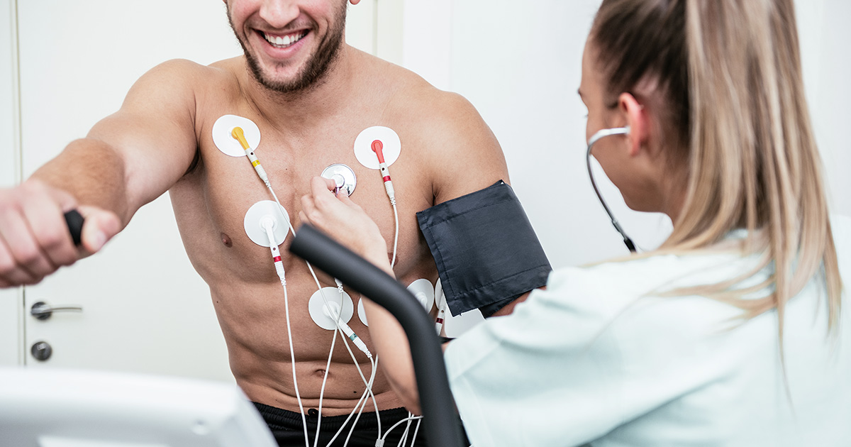
An exercise stress test reveals how the heart works during physical activity. Since exercise stimulates the heart to pump harder and faster, an exercise stress test can identify problems with blood flow within the heart. An exercise stress test usually involves walking on a treadmill or riding a stationary bike with increased intensity, speed and incline while heart rhythm, blood pressure and breathing are monitored and recorded. During the exercise, patients are connected to an electrocardiogram (EKG), allowing heart rates to be monitored. The results obtained from an exercise stress test can determine heart and blood flow problems as well as personal exercise capacity. - Echocardiography (Echo)
An echocardiography uses high frequency sound waves (ultrasound) to create pictures of the heart’s chambers, valves, walls and the blood vessels attached to the heart. The image is called an echocardiogram. During an echocardiography, a probe called a transducer is passed over the chest. The probe produces sound waves that bounce off the heart and echo back to the probe. These waves are changed into images displayed on a video monitor. This test is used to monitor heart structure and its function, allowing the cardiologist to visualize the abnormality of the heart.- Exercise Stress Echocardiography
- Dobutamine Stress Echocardiography
- Transesophageal Echocardiography (TEE)
- Dynamic Electrocardiography (DCG ) or Holter Monitor
A dynamic electrocardiography (DCG) or Holter monitor is a battery-operated portable device that measures and records the electrical activity of the heart continuously over 24 hours. It is used to identify the cause of palpitation or fainting and to diagnose heart arrhythmias. - Vascular Screening Test
Vascular screening test is conducted to detect narrowing or clogged arteries which can potentially lead to the blockage of arteries which supply blood to the heart or brain, causing heart attack and stroke in the future. - Tilt Table Test
A tilt table test involves changing a person’s position quickly (from horizontal to vertical) and determining how blood pressure and heart rate respond. A tilt table test is used to evaluate the cause of unexplained fainting, lightheadedness, dizziness or near-fainting spells whether these symptoms are related to cardiac system.
For more information, please contact
Non-Invasive Cardiology Laboratory
2nd Floor, R-Building, Bangkok Hospital.
Service Hours: Monday - Sunday 07.00 a.m. – 04.00 p.m.