A minimally invasive procedure to treat aortic valve stenosis: Transcatheter aortic valve implantation (TAVI).

Apart from aortic valve regurgitation and rheumatic heart disease, degenerative valvular heart disease is one of the most common and most serious valve disease problems. Recent statistical data reveals that the prevalence of degenerative valvular heart disease has continued rising, especially in the elderly with age-related causes. Other risk factors that aggravate valve degeneration include uncontrolled high blood pressure, diabetes, high cholesterol as well as unhealthy lifestyle e.g. smoking.
Get to know degenerative valvular heart disease
Degeneration of heart valve is typically caused by lipid deposition or calcified nodule formation. Calcium buildup on the valve causes the accumulation of calcium and results in stiffening of the cusps (leaflets) of the valve. This stiffening narrows the aortic valve. As a result, aortic valve becomes more stiff and less flexible with impaired functions of closing and opening. In some cases, if calcium nodules have been formed on the jagged-edge of the valve, it eventually leads to opening failure of the valve which affects the amount of blood supply to the heart. When the blood flow through the aortic valve is reduced or blocked, the heart needs to work harder to pump blood to the entire body while heart wall becomes thicker. Eventually, this extra work limits the amount of blood it can pump, it weakens heart muscle. If it is left untreated, it might cause life-threatening conditions such as acute heart failure.
Degenerative valvular heart disease, particularly “aortic valve stenosis” is serious condition. The aortic valve is located between the lower left heart chamber (left ventricle) and the main artery that delivers blood from the heart to the body (aorta).This heart valve regulates blood flowing in the correct direction. Aortic valve closes off left ventricle that holds the oxygen-rich blood before it is pumped out to the body and opens to allow blood to leave the heart (from the left ventricle to the aorta and on to the body). In aortic valve stenosis, the aortic valve is narrowed , thus the left ventricle has to work harder to pump a sufficient amount of blood into the aorta and onward to the rest of the body. This causes the left ventricle to thicken and enlarge. Eventually the extra work of the heart can significantly lead to heart failure and other heart problems. Aortic valve disease is common in elderly patients, equally in both male and female, with a prevalence of nearly 3% in patients aged 80 years or older. However, with reference to statistical data, male tends to develop this disease approximately 60% while 40% has been presented in female.
Warning signs and symptoms
Signs and symptoms that might indicate the abnormalities of heart valve including valve regurgitation and valve stenosis are:
- Fatigue, especially during times of increased activity
- Feeling faint or dizzy
- Chest pain (angina) or chest tightness with frequency at least 2-3 times per week.

Treatment of degenerative aortic valve disease
Degenerative aortic valve disease cannot be treated with oral medications since cause of this valve condition is derived from valve stenosis (narrowed valve). The ultimate treatment goal is to replace a narrowed aortic valve that fails to open properly. Open-heart surgery, which involves a cut (incision) in the chest, is conventional technique to replace old heart valve with artificial valves, either tissue (biological) valve or mechanical valve. Both types of artificial valves possess their pros and cons. Modern mechanical valves can last extremely long but lifelong treatment with anticoagulants is required, whereas tissue valve does not require the administration of anticoagulants but lifespan is fairly short, compared to the mechanical ones. In the past, to treat aortic valve disease regardless of types of artificial valves, open surgery was essentially needed. Due to the advancements in surgical technology, minimally invasive procedure to treat aortic valve disease without open-heart surgery has emerged. This procedure may be an effective option if the patients are considered to have intermediate or high risk of complications from surgical aortic valve replacement. Conditions that may increase the risk of surgical aortic valve replacement include being the elderly with advanced age and having some underlying diseases such as lung disease or kidney disease.
TAVI: Transcatheter Aortic Valve Implantation
In 1985, Professor Alain Cribier, the Interventional Cardiologist at the Charles Nicolle University Hospital in Rouen, France, performed the first transcatheter aortic valve implantation (TAVI) procedure in the world. He used a PVT percutaneous heart valve without open-heart surgery. TAVI is suitable for patients with aortic valve disease such as aortic valve stenosis. Since it is a minimally invasive procedure, in comparison to open-heart surgery, the advantages of TAVI involve smaller incisions, less blood loss, reduced risk of anesthetic-related side effects, no need the connection of patient with a heart-lung bypass machine during performing surgery, a shorter hospital stay and quicker recovery time which normally requires 2-3 days for hospitalization while it takes up to 7-10 days with open-heart surgery.
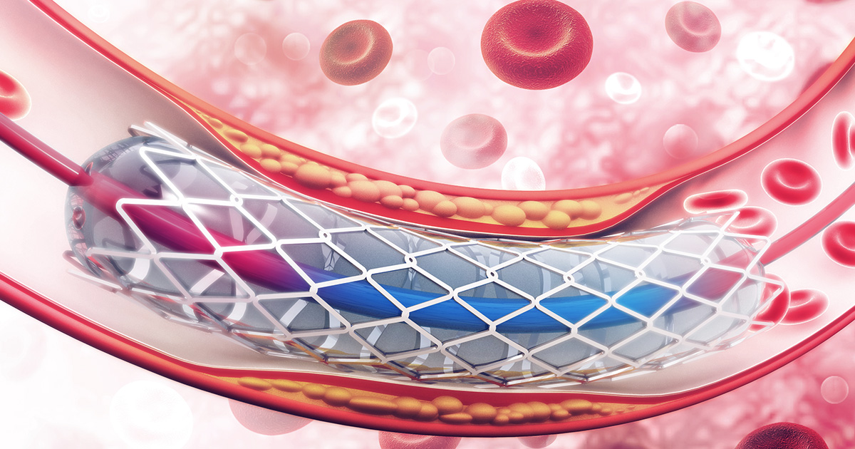
How to perform TAVI
This minimally invasive technique is usually performed through a small cut in the groin and occasionally through a small cut in the side or front of the chest. Tissue (biological) valve is attached to expandable balloon which is further inserted into a 8-10 mm catheter (a thin and flexible tube). Guided by a catheter, this inflatable balloon is first inserted into aorta through a small cut in the groin. With navigation system, after reaching the location of left ventricle and aortic valve, the artificial valve is then placed in position. The inflatable balloon is used to deploy and lock the valve in place. The catheter is further removed and the new valve works in place of the original valve. By performing this procedure, patients have small incisions in the groins, left chest or upper part of right chest, depending on the location of balloon and valve replacement. Currently, TAVI is predominantly considered as an effective treatment option for patients with aortic valve stenosis, not only limited to patients with moderate and high risks.
The most common sites for arterial access are the groin of the leg, shoulder, arm and wrist. The majority of cases has been performed through femoral artery in the groin due to its larger diameter, thus a catheter can be inserted quite easily. However, there are some limitations for artery access through the groin such as circulatory problems in which narrowed arteries reduce blood flow to the legs. In such a case, other sites to reach artery is considerably preferred. Time consumption for TAVI is approximately 2 hours.
Special precautions for TAVI include patients with bacterial infections, patients with acute myocardial infarction, cardiac embolism or blood clot, irregular heartbeat such as tachycardia (rapid heart beat), patients who just recovered from stroke with the administration of anticoagulants and patients with coronary artery disease. After TAVI procedure, patients might need a 3-month anticoagulant therapy. Heavy exercise and physical activities that need extreme exertion must be avoided. Full recovery period normally takes only up to 3 months with an ability to return to daily activities with improved quality of life.